Portfolio
Illuminating the path to new possibilities
Optiscan's groundbreaking solution delivers real-time, non-invasive microscopic imaging at the point of care. With unparalleled clarity and immediacy, our technology facilitates precise clinical decisions, enhances patient outcomes, and streamlines healthcare processes, ushering in a new era of medical excellence.
In today's landscape of escalating healthcare demands and advancing technologies, conventional histopathology - the standard for disease diagnosis, particularly cancer – has its limitations.
Optiscan technology establishes a new standard of precision healthcare through earlier detection, interpretation, and treatment of cancer in the laboratory, operating theatre and beyond.
Unlocking research potential and accelerating new discoveries
Conventional microscopy, enabling examination beyond the human eye's resolution, has been instrumental in deciphering cellular mechanisms in physiology and pathology.
Optiscan's solution allows researchers to visualize living structures in their natural environment, monitor disease progression or treatment, and minimize animal sacrifices. This technology opens new study avenues, elevating research facilities and expediting valuable drug discoveries and therapeutics for human benefit.
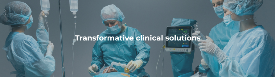
Optiscan's technology facilitates immediate, well-informed clinical decisions and bridges the gap between surgery and pathology. With pathway specific applications, Optiscan is able to enhance patient outcomes through precise, targeted tissue screening and accurate assessment of surgical margins. Serving as a digital enhancement to standard pathology, it achieves slide free biopsy results, ensuring reliability in surgeon diagnoses. Moreover, by reducing the reliance on traditional histopathology and minimizing the need for revision surgery, it brings efficiency gains to healthcare systems.
| Traditional Histopathology: Challenges |
Optiscan Technology: Solution |
| Hours to days to receive results, generating delays in clinical decision making. |
Real-time, in vivo, non-destructive clinical feedback allowing immediate, informed decisions. |
| Single point in time understanding of tissue status. |
Ability to monitor the same area point over time. |
| Incomplete disease identification potential. |
Track treatment impact and potential disease effects. |
| Pathologist and clinician separately located. Potential communication issues. |
Pathologist-clinician collaboration via digital workflow. |
| Samples only a small fraction of possible diseased area. |
Unlimited microscopic sampling across diseased tissues. |
| Single point in time understanding of tissue status. |
Greater economic efficiencies in healthcare systems. |