
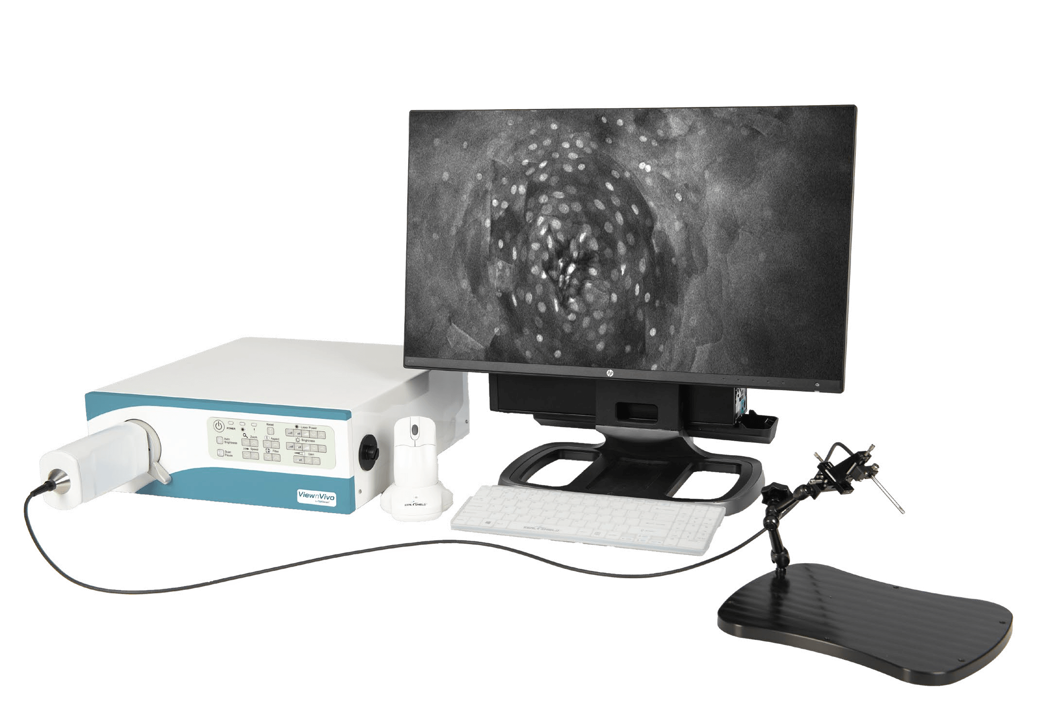
ViewnVivo
See Life Connected
ViewnVivo® offers single-cell level live imaging, providing unique biological system insights and enabling users to see life connected. Developed with our patented confocal imaging technology, ViewnVivo® is a miniaturized in vivo imaging device that offers single-cell 3D live microscopic imaging in the palm of your hand.
Designed specifically for life sciences, pre-clinical and translational research, ViewnVivo® enables users to see systems biology in harmony, maintaining key cell structures, biological systems and cell-to-cell interactions to provide valuable translational results to accelerate drug discovery or unlock new study possibilities.
Why Choose ViewnVivo®?
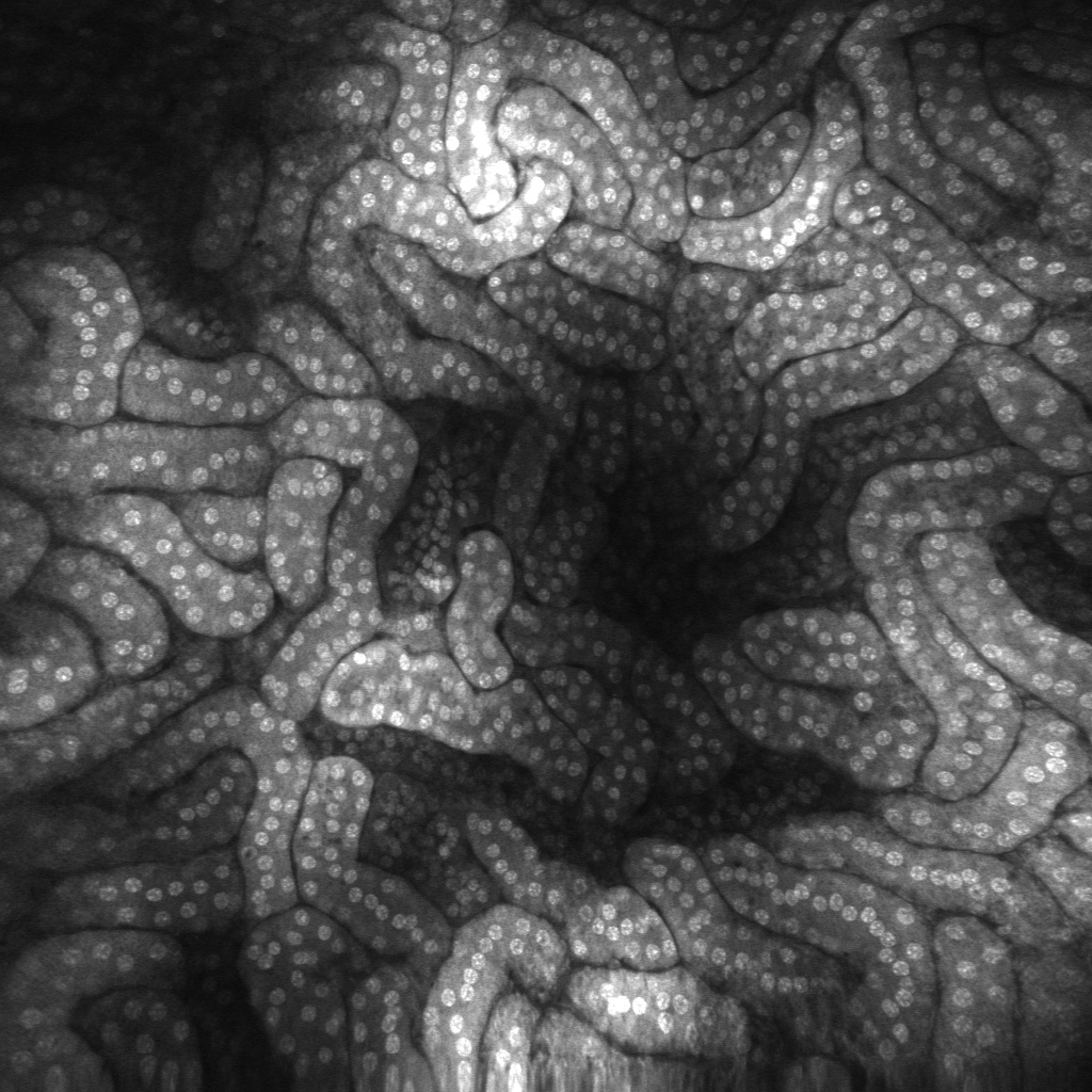 |
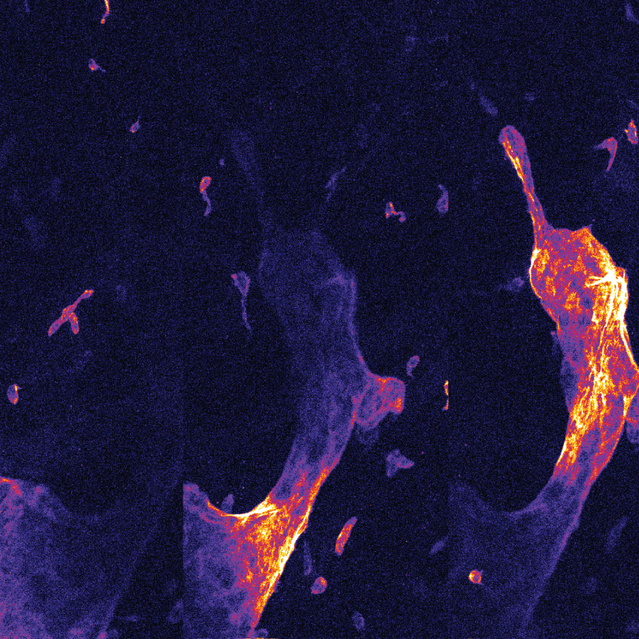
Superb resolution Using a distal end single scanning fibre rather than a proximally placed scanning fibre bundle provides unmatched lateral and axial resolution. The single fibre eliminates dead spaces and broken fibres, delivering high sensitivity and long lifespan. |
 |
Never miss an image The ViewnVivo® imaging software includes an Image Rollback function. Users can select and save images from up to 60 memory held frames. Individual images or image selections can be saved or made into movie files. |
 |
Longitudinal studies High level disinfection and sterilization options provide a cost-effective, practical way to enable longitudinal studies on the same subject, as well as the ability to image cell cultures and other samples where sterility is required. |
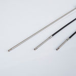 |
Long-life probes ViewnVivo® probes are not a consumable item and will retain their imaging quality through years of operation and hundreds of procedures. |
 |
Precise control in the Z-dimension ViewnVivo® reports the position of the focal plane with micron accuracy. The imaging software provides an intuitive interface to set and collect Z-Stacks. This data can be turned into movies or ported into Fiji for processing. |
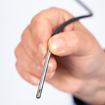 |
Image from any angle The handheld flexible probe allows the user access to previously inaccessible samples. |
ViewnVivo® Benefits:
- Slide-free and biopsy free
Users can generate images at the single-cell level, in real-time. - Viewing live biological systems in-vivo at the cellular level in their entirety
Not achievable through other imaging techniques. - Non-destructive
Image creation is non-destructive to the tissue of interest. - Portable
Easy to work on live tissues. - Image depth variation
Unique ability to vary image depth in the Z-Axis: up to 400 microns (tissue dependent). - Zero cost image acquisition
Just staining cost required. - Post-processing versatility
Images can be easily post-processed to produce a range of presentations (incl 3D).
Potential Applications:
| Cancer research |
|
Tissue regeneration |
|
| Cell tracking |
|
Photodynamic Therapy (PDT) |
|
| Infection & treatment |
|
Pharmacology |
|
| Pathology |
|
Animal models |
|
| Microvascular research |
|
Molecular Imaging |
|